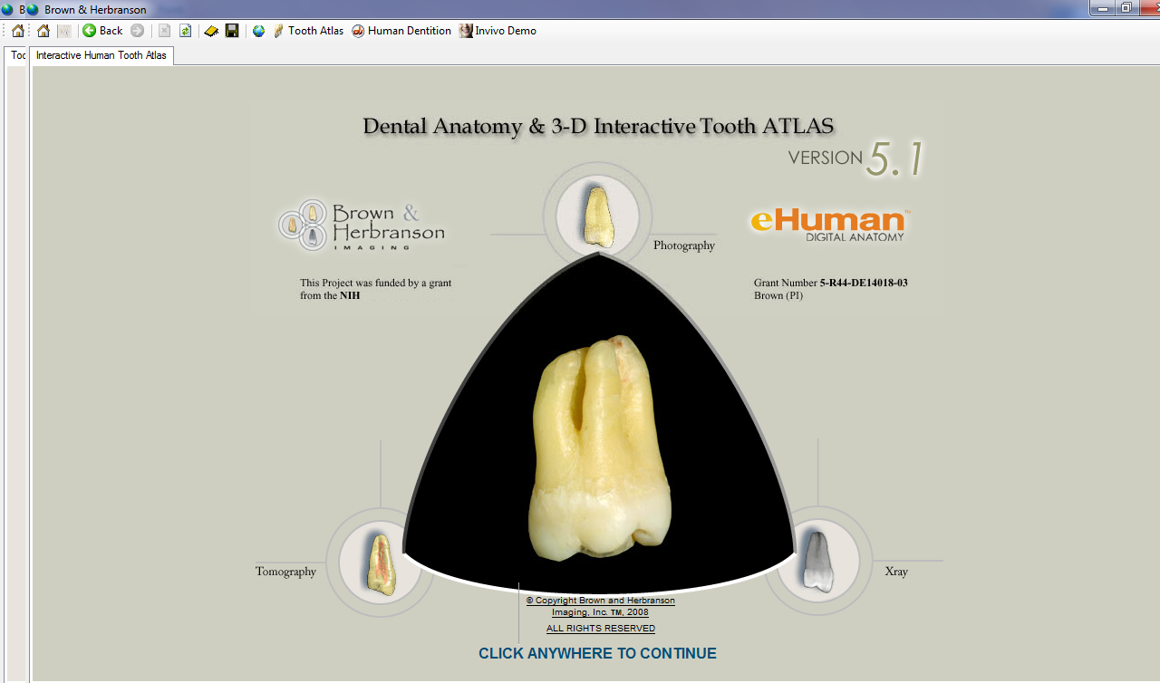
Dental Anatomy & 3D Interactive Tooth Atlas
Year: 2008
Version: Version 5.1
Platform: PC (Vista, XP, Win2000)
Compatibility with Vista: complete
System requirements: 256 MB RAM
32 MB - 64 MB graphics card
2 GB of free harddrive space
1024x768 monitor resolution minimum
1.5 GHz processor or higher
Language: English
Medicine: Present
Description
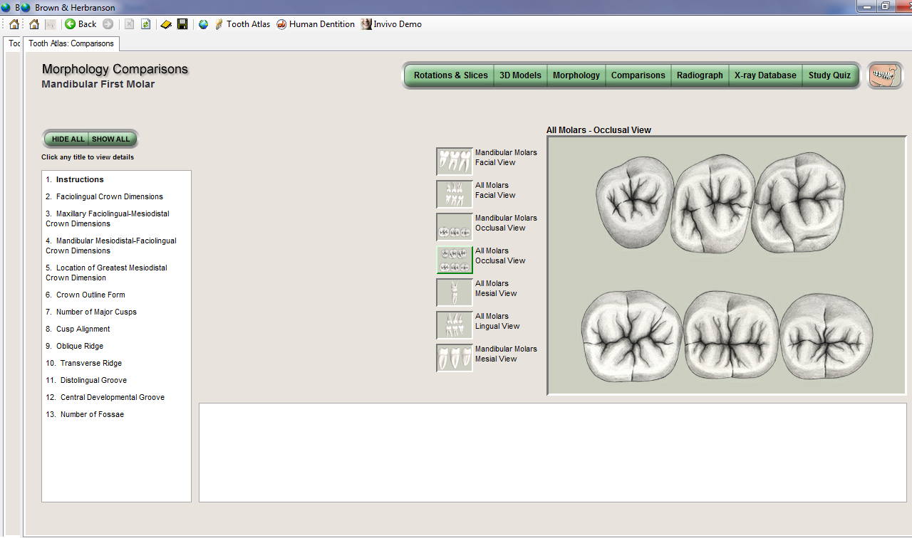
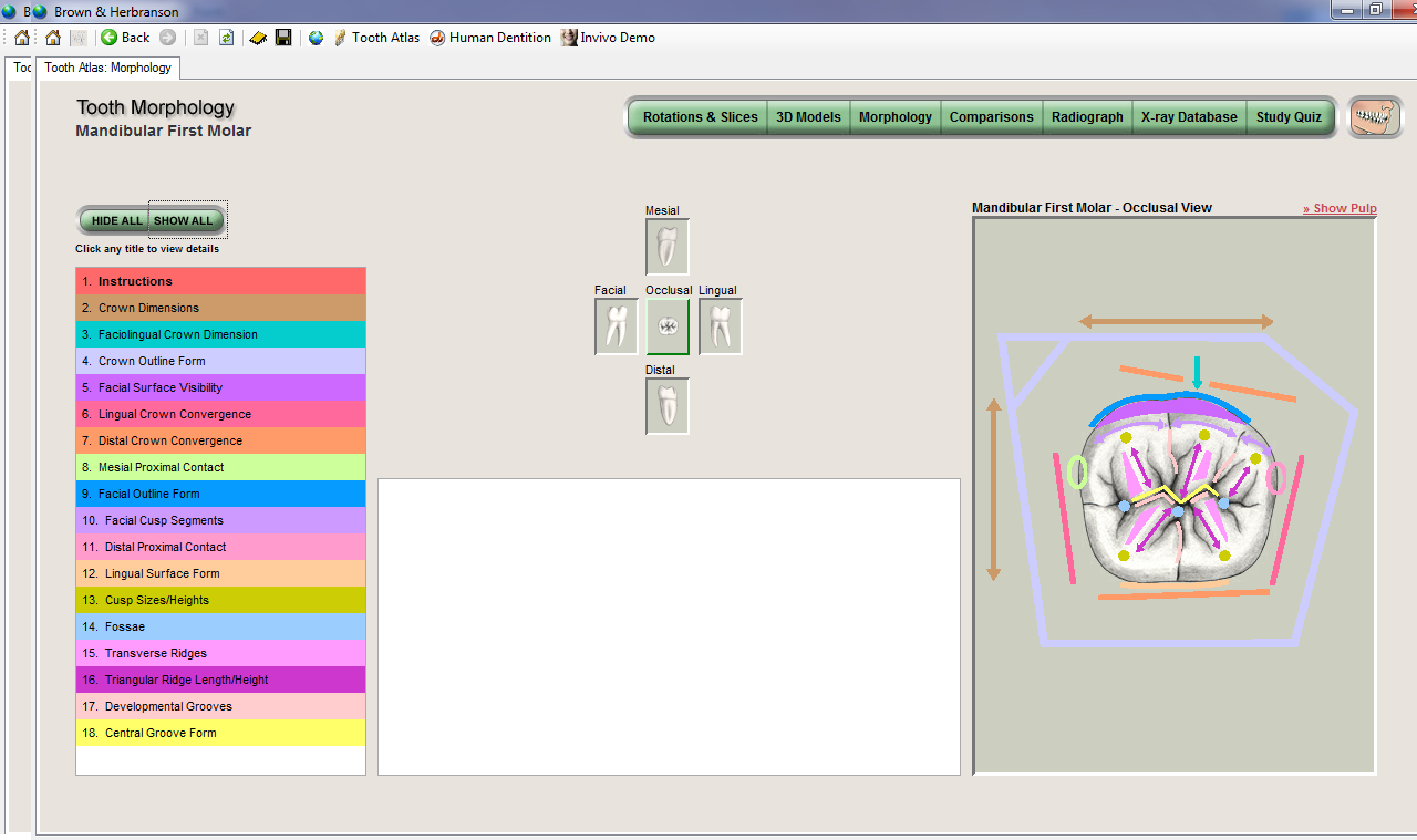
Recently, three-dimensional X-ray microtomography has been available for researchers thanks to the emergence of a number of commercial systems for the study of small animal models. Just like a traditional computer tomography was used to create a detailed anatomical atlas of the entire human body, X-ray microtomography has proved a valuable and effective tool for visualization of human teeth. To our knowledge, a comprehensive atlas of comparable data on human teeth there. This database has been collected under controlled experimental conditions, thanks to which each tooth was used the same method of operation. This atlas is a valuable textbook for researchers studying the morphology of human teeth. Base mikrotomograficheskih data serve as a valuable source of information for researchers dealing with morphology and mineralization of teeth
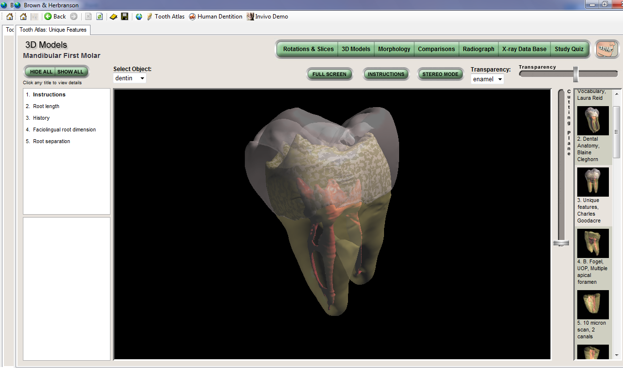
Anatomy of the apical third of the root canal is of particular interest for endodontists, because it affects the nature of the treatment procedure. The apical third of the canal may be a plurality of branches extending in different directions. Fig. 12, a, b is represented by a root canal, having a number of branches. One of the objectives of the "Atlas of teeth" is a demonstration of common varieties of root canal anatomy
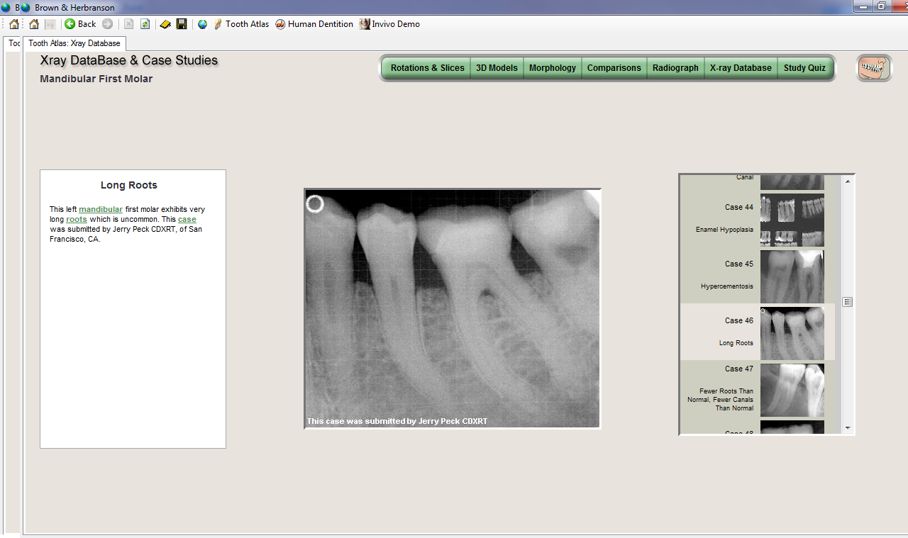
Extras. Information: The study of a large number of teeth made it possible to identify not as widespread anomalies. Typically, the root of the tooth is covered with a thin layer of cement. When a phenomenon known as gipertsementoz, on top of the primary layer of cement produced excessive amounts of cement. Fig. 13 shows mikrotomografichesky section taken along the axis of the root, which can be seen the line, showing a normal cement layer and excess cement thickness of about 1.5 mm
Version: Version 5.1
Platform: PC (Vista, XP, Win2000)
Compatibility with Vista: complete
System requirements: 256 MB RAM
32 MB - 64 MB graphics card
2 GB of free harddrive space
1024x768 monitor resolution minimum
1.5 GHz processor or higher
Language: English
Medicine: Present
Description
Recently, three-dimensional X-ray microtomography has been available for researchers thanks to the emergence of a number of commercial systems for the study of small animal models. Just like a traditional computer tomography was used to create a detailed anatomical atlas of the entire human body, X-ray microtomography has proved a valuable and effective tool for visualization of human teeth. To our knowledge, a comprehensive atlas of comparable data on human teeth there. This database has been collected under controlled experimental conditions, thanks to which each tooth was used the same method of operation. This atlas is a valuable textbook for researchers studying the morphology of human teeth. Base mikrotomograficheskih data serve as a valuable source of information for researchers dealing with morphology and mineralization of teeth
Anatomy of the apical third of the root canal is of particular interest for endodontists, because it affects the nature of the treatment procedure. The apical third of the canal may be a plurality of branches extending in different directions. Fig. 12, a, b is represented by a root canal, having a number of branches. One of the objectives of the "Atlas of teeth" is a demonstration of common varieties of root canal anatomy
Extras. Information: The study of a large number of teeth made it possible to identify not as widespread anomalies. Typically, the root of the tooth is covered with a thin layer of cement. When a phenomenon known as gipertsementoz, on top of the primary layer of cement produced excessive amounts of cement. Fig. 13 shows mikrotomografichesky section taken along the axis of the root, which can be seen the line, showing a normal cement layer and excess cement thickness of about 1.5 mm
2.28GB